Synovial Fluid
Instructor:
Mark H. Wener, MD; Adam R. Orkand, BA; Michael L. Astion, MD, PhD
Contact Hours:
1.0
Date:
January 01, 2025 - December 31, 2026
Description:
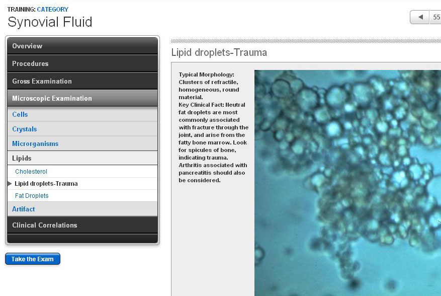
Classification of synovial effusions. Gross and microscopic examination, including the use of polarized light.
Learning Objectives:
- Describe the basic classification of synovial effusions.
- Describe the gross appearance of synovial fluid specimens.
- Discuss the principles and applications of polarizing microscopy and compensated polarizing microscopy, and how to perform them.
- Distinguish urate crystals from calcium pyrophosphate dihydrate (CPPD) crystals.
- Describe other crystals, artifacts, inclusions, and cell types that may be present in synovial fluids.